- February 11, 2022
Kyla learned how cultural differences, even within Western culture, equip us to accomplish more together than individually.
Kyla Truman created an image atlas of brain slices for an Alzheimer's Disease brain originally thought to be neurotypical.
Incredible collaboration
"Through MISTI, I was able to learn about the incredibly collaborative aspects of neuroscience research, as well as how findings are communicated internationally. My time in Madrid taught me a great deal about cultural differences, even within Western culture, and how these differences equip us to accomplish more together than individually," remarks recent Brain and Cognitive Sciences grad, Kyla Truman. While many seniors found themselves whisked away to exotic vacation spots to celebrate Commencement, Kyla spent her time in a small lab at Instituto Cajal in Madrid, Spain and had an unforgettable experience.
Neurolucida and Imaris
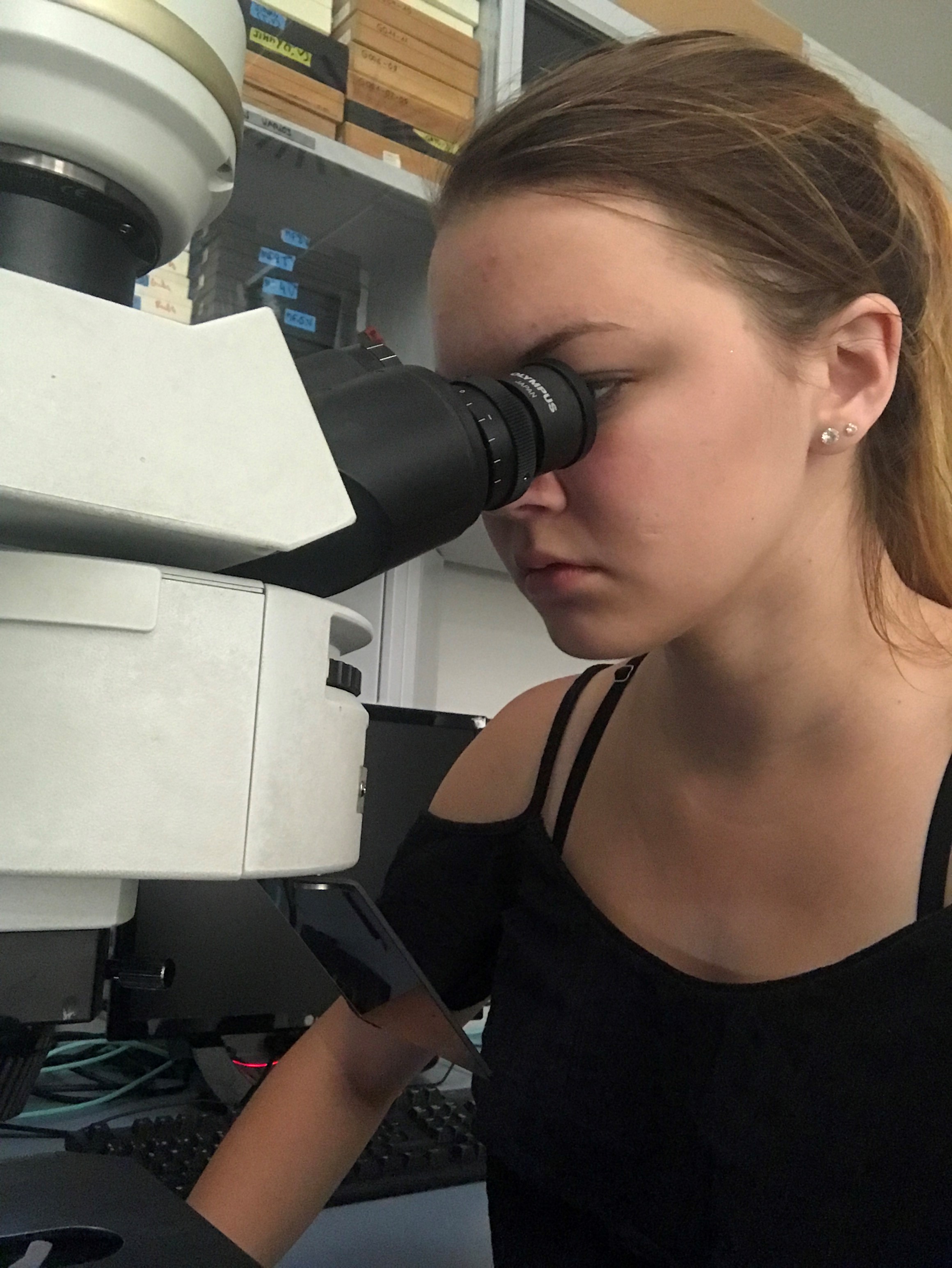
While at Instituto Cajal, Kyla adjusted well to the flexible Spanish schedule and enjoyed going to lab in the early afternoons. Upon arrival by bus, she would gather her samples and sit down to the microscope or computer to begin her projects. The first project she contributed to was a comparison of two 3D neuronal reconstruction softwares, Neurolucida and Imaris. Kyla examined a cortical pyramidal neuron, known for having extensive dendrites. While tracing the soma and dendrites was fairly autonomous, Kyla spent the majority of her time reconstructing dendritic spines, paying close attention to the shape of the neck and head of each spine. Each neuron had previously been injected and immunostained with anti-Lucifer yellow antibody, making it easy to view and trace. Neurolucida software was easier to manipulate and user-friendly, but the tracings of the dendritic spines were not as exact as they were with the more tedious Imaris software.
Seeing an invisible disease
After completion of this software comparison, Kyla began a new project focused on creating an image atlas of brain slices from a brain previously believed to be neurotypical control. Upon examination under high magnification microscope, however, cells appeared positive for Tau protein, which is closely related to the debilitating symptoms seen in Alzheimer’s Disease (AD). The brain, provided by donation from a hospital, turned out to actually have AD. Kyla took hundreds of photos of the brain tissue, which can be stitched together to form full images of brain areas that had the most Tau protein, such as the hippocampus. The distribution of these Tau-positive cells may lend insight into how AD develops and spreads through the brain. “It was extraordinary to work with human brain samples and see with my own eyes how a typically ‘invisible’ disease develops,” noted Kyla.